Digital & Innovation
Harnessing the power of artificial intelligence in pathology to detect cancer
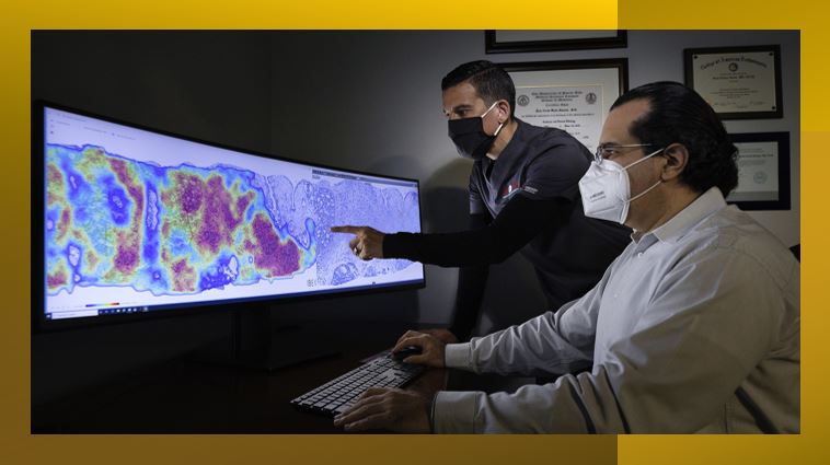
Digital & Innovation: A large international collaboration, led by University of Sydney, has developed an innovative, advanced artificial intelligence (AI) application, PathoFusion, that could be used for the examination of routine tissue samples in order to identify indications of cancer.
The research melds contributions from computer scientists, neuropathologists, neuosurgeons, medical oncologists and medical imaging scientists.
ANSTO’s Prof Richard Banati, a Professor of Medical Radiation Sciences/Medical Imaging, who studies the brain’s innate immune system using advanced medical imaging techniques, is a co-author on the paper published in the journal, Cancers.
“The idea behind PathoFusion was to create a novel advanced deep learning model to recognise malignant features and immune response markers, independent of human intervention, and map them simultaneously in a digital image,” explained Banati.
The experiment to evaluate the model involved the examination of tissue from cases of glioblastoma, an aggressive cancer that affects the brain or spine.
The team used the expert input of neuropathologists to ‘train’ the software to mark key features.
Experiments confirmed that the application achieved a high level of accuracy in recognising and mapping six typical neuropathological features that are markers of a malignancy.
Pathofusion reliably identified forms and structural features with a precision of 94% and sensitivity of 94.7% and immune markers at a precision of 96.2% and sensitivity of 96.1%.
The application combines layers of information about dead or dying tissue, the proliferation of microscopic blood vessels and other vasculature with the expression of a tumour genetic marker, CD276, in an image that combines the data in a heatmap.
“The research confirmed that it is possible to train neural networks effectively using only a relatively small number of cases, that should be useful for some scenarios,” said Banati.
Painstaking and time-consuming routine morphological diagnostic work is carried out by pathologists, who examine individual slides under a microscope to mark and quantify features that are markers of disease and provide the information to clinicians.
“Anticipating further hardware improvements in computing, it should exceed the speed of human microscopic feature recognition by orders of magnitude when whole histological slides are used at high resolution,” said Prof Manuel Graeber of the University of Sydney and Brain and Mind Centre.
“In future, the model could improve the workflow of a neuropathology or pathology unit by facilitating microscopic analyses or benefit patients in area without local access to these services, “ explained Banati.
Contributors to the research included The University of Sydney, The Brain and Mind Centre, Xuanwu Hospital (China), St Vincent’s Hospital, Cooperative Trials Group of Neuro-Oncology, Brain Cancer Consultancy and University of Western Australia.
News & Trends - Biotechnology

CSL reshapes R&D while bracing for U.S. tariffs
Australia’s largest biotech company CSL is streamlining its R&D operations to enhance efficiency amidst a rapidly evolving global landscape. The […]
MoreNews & Trends - MedTech & Diagnostics
Australia joins Medtronic trial in fight against resistant hypertension
Medtronic has launched an international clinical trial across Australia, the United States, and Europe to evaluate the feasibility of multi-organ […]
MoreNews & Trends - MedTech & Diagnostics

Medibank launches pharmacogenetic testing while government stalls on insurance discrimination ban
Medibank has become the first Australian health insurer to pay towards pharmacogenetic testing (PGx) for eligible customers on Extras cover. […]
MoreNews & Trends - Pharmaceuticals

Global pledge shifts visibility and action for patients with advanced breast cancer
Three breast cancer organisations have united internationally to demand that people living with metastatic breast cancer (MBC) are no longer […]
More